Diabetic Foot Ulcers

Diabetic foot ulcers are one of the major complications of diabetes. Between 19 and 34% of all diabetics will develop a foot ulcer at some point in their lives (1). Often underestimated, or even unrecognised by the patients affected, it can lead to complications such as an infection requiring hospitalisation, or even in the worst cases, amputation. After amputation, patient life expectancy does not exceed 5 years in almost 70% of cases, which is lower than that for certain types of cancer (2). This outcome is not however a fatality. With proper prevention, ulcers can be avoided.

What causes diabetic foot ulcers?


Diabetes does not only cause blood sugar imbalance. It also causes two phenomena which increase the risk of occurrence of a diabetic foot ulcer:

  • Involvement of the distal nerves of the legs, especially with loss of sensitivity. As a result, the patients only rarely feel they have an ulcer (neuropathic ulcer).
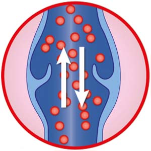
  • Blocked arteries, ultimately leading to insufficient blood flow (ischemic ulcer).

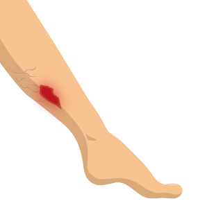
In three out of four cases, diabetic foot ulcers are related to wearing uncomfortable shoes, to an injury caused when cutting the toe nails, to a burn/friction or to fissures / cracks on the soles of the feet.

The longer an ulcer remains open, the more the risk of infection increases. It can lead to amputation in extreme cases.

What is a diabetic foot ulcer?

Plaie du diabétique définition


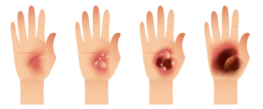
Caused by an initially minor lesion (a simple cut through to wearing uncomfortable shoes), a diabetic foot ulcer can take three forms:

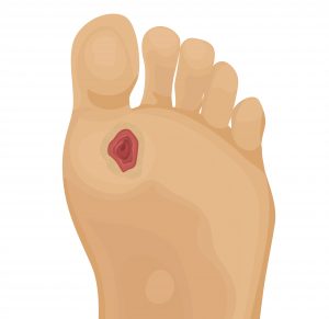
  • Neuropathic ulcer. Appearing on the toes or under the arch of the foot, as a perforating ulcer of the foot, on the foot joint, this ulcer begins with a callus or corn. It comes with a loss of sensitivity (pain/heat), has clear edges and the skin is dry. In effect, diabetes causes neuropathy which affects the sensory and motor nerves, weakening the extremities of the lower limbs. Therefore the patient’s foot becomes deformed and their nails become curved like claws. The loss of sensation in the foot can even prevent the patient feeling they have an ulcer, which can then rapidly become infected.
  • Ischemic ulcer. This is related to a perfusion defect. It mainly occurs on the tips of the toes, on the heel or edge of the foot. The skin breaks, and is pale or bluish in colour. The foot is cold and painful, and the patient can feel burning or itching sensations. Yellow, red, pink, or even black if it is necrotic, the ulcer can take on different colours, depending on its stage of progression.
  • Neuroischemic ulcer. It is related to neuropathy and is associated with The ulcer forms on the edge of the foot or tips of the toes, or even under the toenails. The first sign is generally a blister caused by repeated friction of the foot in shoes that are too tight or too narrow.

In adults, an infection is likely to occur in addition to the ulcer. In effect, the open wound is subject to bacterial invasion. Diabetic foot ulcer therefore makes diabetes the leading cause of amputation in the world3.

Diabetic foot ulcer prevention

The severity of the foot involvement requires targeted prevention according to the risk. The Haute Autorité de Santé (HAS)4 has therefore defined a podiatric risk grading system in order to adapt treatment. The foot assessment is used to grade the level of risk according to the following classification:

Grade 0: no sensory neuropathy

Podiatric risk grade 0 means there is no loss of sensitivity in the feet. Therefore, the risk is the same as in the general population. It is the most common situation. Standard hygiene rules should be followed, like wearing appropriate, non-traumatic footwear or avoiding maceration (feet should be dried properly and synthetic socks should be avoided) and traumatic treatments (e.g.: use of a scalpel). It is especially important to balance the diabetes as far as possible and to control any risk factors (e.g.: smoking, arterial hypertension, cholesterol etc.).

Grade 1: isolated sensory neuropathy

In addition to the recommendations that apply for grade 0, you can also follow the advice below:

  • If the ulcer is very painful or uncomfortable, talk to your doctor about it. Certain medicines can provide relief, in addition to balancing blood sugar.

  • If the ulcer involves loss of sensation, you or a member of your family must inspect your feet daily. It will enable you to detect any ulcers that go unnoticed. It is essential to see a doctor as soon as possible if there is an ulcer, even if it is not painful.

It is especially important to not damage the feet using too abrasive and/or corrosive treatments, by wearing shoes that hurt or walking barefoot etc. Apply a moisturising cream daily if your feet are very dry. Of course, your diabetes should be controlled as effectively as possible to prevent the neuropathy worsening.

Grade 2: sensory neuropathy with arteriopathy of the lower limbs and/or foot deformation

Grade 3: history of ulcer or amputation

These very high risk feet (grade 2 and grade 3) require both increased vigilance on your part and/or by your family, but also active ulcer prevention measures. Podiatrists or nurses can remove the hyperkeratosis (callus). Podiatrists/chiropodists/orthotists can make ortheses or insoles to correct abnormal weight bearing areas. Sometimes, if your foot is very deformed, you may even have to wear specially-made shoes. If there is arterial involvement, revascularisation may be essential.


Good to know !

Two types of prevention can be paid for on prescription for diabetic patients with podiatric risk grade 2 or 3:

  • a yearly preventive treatment for grade 2 risk foot lesions. It includes 4 prevention sessions per year maximum;
  • a yearly preventive treatment for grade 3 risk foot lesions. It includes 6 prevention sessions per year maximum.

The prevention treatments dispensed at the patient’s home must come with a medical prescription to be reimbursed.

The yearly prevention package includes an initial foot assessment, prevention sessions and a brochure.

Treatment

First of all, diabetic foot ulcers require rapid and comprehensive treatment to minimise the risk of complications. Regardless of its size, the ulcer must then be closely monitored by a multi-disciplinary team. The team includes the diabetes specialist, the general practitioner, the nurse, the podiatrist-chiropodist and the surgeon who will all closely monitor the more or less rapid progress of the ulcer.

Wearing a pressure relief device is also compulsory. It means the foot does not press on the ulcer and more effectively distributes pressure on walking, so as not to aggravate the wound.

It is essential to apply a dressing after careful ulcer cleansing and removal of denervated tissue. Its aim is to promote healing.

The diabetes, comorbidities and nutritional status must also be managed at the same time.

And afterwards?

70% of healed diabetic foot ulcers relapse within 5 years5 . Diabetic patients must therefore take special care of this part of their body, by following some basic recommendations:

  • Wear shoes suited to the shape of the foot, and always with socks. 
  • Never walk barefoot
  • Show their feet to a chiropodist on a regular basis
  • Monitor the general foot condition (use a mirror to inspect the sole)
  • Continue to closely monitor blood sugar levels and follow a diabetes-reducing diet.
  • Ask a podiatrist or chiropodist to look after their feet and nails (cutting/filing) and not use corn remover (product to remove corns)
  • Wash their feet daily with soap and water, not forgetting to dry between the toes and user a moisturiser to keep them supple.
  • Not warm their feet using a heat source (e.g. Hot water bottle)
 
1- Armstrong DG, Boulton AJM, Bus SA. Diabetic foot ulcers and their recurrence. N Engl J Med 2017; 376: 2367-75
2- Walsh JW, Hoffstad OJ, Sullivan MO, Margolis DJ. Association of diabetic foot ulcer and death in a population-based cohort from the United Kingdom. Diabet Med 2016; 33:1493–98.
3- Whiting, D. R., Guariguata, L., Weil, C., and Shaw, J. 2011. “IDF Diabetes Atlas: Global Estimates of the Prevalence of Diabetes for 2011 and 2030.” Diabetes Res. Clin. Pract. 94 (3): 311-21.
4- Evaluation of the acts performed by the pedicure-podiatrist for the prevention of grade 1 risk foot injuries in the diabetic patient. French Health Authority. (Évaluation des actes réalisés par le pédicure-podologue pour la prévention des lésions des pieds à risque de grade 1 chez le patient diabétique. Haute Autorité de Santé)
5- Connor H, Mahdi OZ. Repetitive ulceration in neuropathic patients. Diabete
Everyone living with diabetes should have a foot check by a health care professional at least once a year. If you’ve not had your foot check this year, ask your podiatrist, GP or nurse for one.
Go to your doctor or podiatrist straight away if you notice anything unusual or worrying during your foot check such as:
  • an ulcer
  • a scratch
  • a cut
  • a blister
  • feel pain
  • swelling
  • redness

Treatment of diabetic foot ulcers

It’s really important to contact you GP, podiatrist or nurse immediately if you see something wrong – no matter how small the wound.

The important thing to remember is to keep your weight off your foot.

A key factor to close the wound and to avoid complications such as infection and amputation is to react early. It is important that your feet are being taken care of by a team of specialists

For more information on Diabetic Foot Ulcer please visit Save Feet Save Lives

1. Setacci C, de Donato G, Setacci F, Chisci E. Diabetic patients: epidemiology and global impact. J Cardiovasc Surg (Torino). 2009 Jul, 50(3) : 263-73
 
2. Armstrong DG, Boulton AJM, Bus SA. Diabetic foot ulcers and their recurrence. N Engl J Med 2017; 376: 2367-75
 

Leg Ulcers

What is a leg ulcer ?

Leg ulcers are chronic wounds located on the lower thirds of the leg (under the knee). The most common cause is poor blood circulation, particularly the inability of the veins to return deoxygenated blood from the legs back to the heart. Other causes or risk factors include prolonged pressure on an area (such as long-term lying in bed in one position), poorly managed diabetes, high cholesterol, smoking, dietary problems and poor arterial circulation.

Leg ulcers can be:

  • Venous leg ulcers : related to poor venous circulation. The wound is superficial in most cases, with inflamed, uneven edges. It is painful and mainly located around the ankle.
  • Arterial leg ulcers : related to poor arterial circulation. These wounds may start out as a small cut with uneven edges on the leg or foot/toes and may progressively become deeper. These wounds are usually very painful.
  • Mixed leg ulcers: related to poor arterial and venous circulations. Signs of a venous ulcer and an arterial ulcer are present at the same time.

How does a venous ulcer form?

A venous leg ulcer is the sign of chronic venous insufficiency which prevents the blood flowing normally in the veins of the leg. When blood flow is blocked, the blood stagnates in the leg veins causing tissue damage and skin breakdown.    

Normal Vein

The veins allow blood to return to the heart. When you walk or exercise muscles contract to pump blood in the right direction. One-way valves stop blood from travelling backwards in the vein.

Damaged Vein

When veins are damaged the one-way valves that stop blood from travelling backwards in the vein stop working. The pooling of blood stretches and distorts the vein. This results in damage to the tissue surrounding the vein – the skin is no longer correctly oxygenated and becomes fragile. There is high risk of an ulcer forming from slight trauma or even spontaneously.


Various factors can increase the risk of chronic leg ulcerations, including:

  • History of varicose veins or deep vein thrombosis
  • Age
  • Arterial disease
  • Cigarette smoking
  • Being overweight/obesity
  • Lack of physical exercise or bed-bound

Treatment of venous leg ulcers

It is very important to consult your doctor as soon as you notice a leg ulcer. Treatment includes:

  • Cleaning the wound to remove the dead tissue
  • Specialised dressings – dressing are changed less often these days, because frequent dressing changes remove healthy cells as well.
  • Occlusive (air and water-tight) dressings are often used as ulcers heal better when they are covered.
  • Compression treatment – boosts internal pressure, using either elasticised bandages or stockings. This is particularly effective if multiple layers are used.

For a mixed leg ulcer (both venous and arterial), compression can be used but it is looser than for a venous ulcer. Compression treatment is not used for arterial ulcers.

Avoiding relapse

To reduce the risk of developing other ulcers you should make sure any underlying conditions that contributed to your leg ulcer are addressed and treated. These may include:

  • treatment for varicose veins
  • quitting cigarettes
  • improving our diet
  • taking regular exercise

You should also:

  • avoid crossing your legs whilst sitting down
  • avoid hot baths
  • keep you leg elevated above the level of your heart where practical
  • wear your compression socks
  • wear comfortable, non-slip shoes
  • regularly moisturise your skin
  • prevent weight gain
 
1. Victorian Government Department of Health. Better Health Channel. Leg ulcers. Available at: http://www.betterhealth.vic.gov.au/health/conditionsandtreatments/leg-ulcers

Pressure Injuries

What is a pressure ulcer?

A pressure ulcer is a chronic wound which appears when sitting or lying for a prolonged period which creates pressure in the weight-bearing areas. This pressure compresses the tissue between two hard surfaces prevents the blood from flowing normally leading to reduced oxygen supply, which causes damage to the tissue and where the wound eventually develops sometimes in just a few hours.

In 80% of cases, a pressure ulcer appears on the sacrum or the heel, mean weight-bearing areas in disabled patients or patients with reduced mobility. The wound can also occur on the elbows, shoulder blades, or the back of the head.

Pressure ulcers are classified by stage according to their severity.

  • Stage 1 : Persistent redness. Stage 1 pressure ulcer is redness which appears and which does not disappear when pressed. . The skin is not yet broken, and there is not yet a wound. Specific treatment must be started at this stage. In this case it is recommended to not massage the area as this may aggravate the condition.
  • Stage 2 : Erosion of the skin of the epidermis and the dermis. The wound is formed and the skin is broken.
  • Stage 3 : The epidermis, the dermis and the hypodermis are affected. There is necrotic (black) tissue and scabs. NB: the pressure ulcers always appears smaller on the surface than it is deeper down.
  • Stage 4 : Deep pressure ulcer. Deep necrosis with possible involvement of the bones, tendons and muscles.

Factors promoting the appearance of pressure ulcers:

  • Immobility
  • Patient age. Even if pressure ulcers can also occur in children, patients over the age of 70 are especially at risk.
  • Malnutrition
  • The quality of the skin. The finer it is, the more sensitivities to friction, to shear stress and to pressure.
  • Incontinence, which causes permanent humidity and renders the tissue or fragile due to maceration.
  • Impaired motricity and lack of mobilisation
  • Change of sensitivity. Patient (sometimes with neurological disorders) does not feel the pain and does not have the reflex to change position.

Pressure ulcer treatment

Regardless of the stage of the pressure ulcer, treatment must be started as soon as possible.

The type of treatment depends on the stage of the pressure ulcer (reversible or irreversible lesion) what is in most cases synonymous with healing.

If there is a visible lesion, it should be protected by the appropriate dressing.

Where both the epidermis and the dermis are involved, the nursing staff will look after wound cleansing and healing.

Regardless of the severity of pressure ulcer first thing to do is to remove pressure. Patients position should be changed every 2 to 3 hours.

Setting up a specialised bed, specific mattress (foam, water, air) and cushions, will relieve and distribute pressure in the high-risk areas.

Treatment should come with appropriate nutritional management and management of the various comorbidities.

And afterwards?

When we know that two hours without moving is enough to create a pressure ulcer the following prevention measures will be just as useful to the patient as to their carers:

  • Always examine the areas of the body in which a pressure ulcers is likely to develop (weight-bearing areas).
  • Move the patient, even if it means calling on a nurse or physiotherapist.
  • Fully wash the patient at least once a day and clean the perineal area each time the patient goes to the toilet.
  • Avoid using rough fabric to dry the skin.
  • Change sheets daily.
(1) Quoted by C. Revaux in the Consensus Conference Prevention and Treatment of Pressure ulcers in Adults and the Elderly (Conférence de consensus Prévention et traitement des escarres de l’adulte et du sujet âgé). 15/16 Nov 2001. Georges Pompidou European hospital

Burns


Contact with hot drinks, food, fats and cooking oils was the most common cause of thermal injuries however explosions, chemical burns, contact with fire, smoke or flames and exposure to light and radiations can also cause burns. All burns require immediate first aid treatment. Burns can lead to functional, aesthetic and psychological effects. If there is significant skin loss there is risk of infection.

What is a burn?

A burn is a skin lesion which sometimes affects underlying tissue. There are four types:

  • Thermal – occurs due to contact with a solid (eg embers, hot iron, oven), liquid (boiling water/oil/burning hydrocarbon), or an explosion. Biting cold can also cause burns.
  • Chemical – caused by a caustic product (eg. acid)
  • Electrical – caused by contact with an electric current or arc.
  • Radiation – caused by solar UV radiation, XRay or nuclear radiation.

Types of burns

The severity of a skin3 depends on three main criteria: depth of burn, extent over the body and location on the body.

How is the depth of a burn evaluated?

There are three levels of burns:4

  • Superficial – these burns cause damage to the first or top layer of skin only. The burn site will be red and painful.
  • Partial thickness – these burns cause damage to the first and second skin layers. The burn site will be red, peeling, blistered and swelling with clear or yellow-coloured fluid leaking from the skin. The burn site is very painful.
  • Full thickness – involves damage to both the first and second skin layers, plus the underlying tissue. The burn site generally appears black or charred with white exposed fatty tissue. Very deep burns may damage the underlying muscle or bone. The nerve endings are generally destroyed and so there is little or no pain at the site of the full-thickness burn. However, surrounding partial thickness burns will be very painful.


Les degrés de la brulure


It can be difficult to tell the difference between partial and full-thickness burns. The depth of a burn is not critical in the initial treatment of burns. An assessment of the extent of the burn is more important initially.


How is the extent of a burn evaluated?

The extent of burn is estimated as a percentage of total body surface area.

Location

Burns to some areas such as the face, hands, feet and perineum are taken particularly seriously.

Other factors to be considered include the person’s age (children under 5 or adults over 60) and coexisting chronic diseases (eg heart failure, respiratory failure, diabetes).

First Aid for burns

Remove the person from danger and further injury. Hold the burn under cold running water for 20 minutes. If necessary, prevent heat loss by covering unburnt areas.

Burnt clothing should only be removed if it does not stick to the burn. Do not remove clothing that is stuck to the burn as this carried a risk of skin detachment. Any chemicals, such as acids and alkalis, must be washed off with running water for at least 20 minutes. A cool shower is ideal.

Superficial burns required pain relief, dressings and regular review to make sure they don’t become infected.

A major burn is defined as a burn of any depth that involves more than 20% of the total body surface area for an adult (and more than 10% of total body surface area for a child).

Major burns are a medical emergency and require urgent treatment. Immediately apply cold water to all affected areas and then cal triple zero (000) for an ambulance. A cool or lukewarm shower is ideal.

Partial thickness burns will require a non adherent dressing to promote healing and reduce the risk of infection. If the body is not able to heal the injury by itself, skin grafts may be needed.


(1) Australian Government. AIHW. injury in Australia: burns and other thermal causes.Updated 10 Mar 2020 Available at: http://www.aihw.gov.au/reports/injury/burns-and-other-thermal-causes
(2) Annual report on burns epidemiology in France. 2008. http://www.sfb-brulure.com/index.php/documentation/epidemiologie.html
(3) Assurance Maladie. How to rapidly assess the severity of a burn? Website Ameli-sante.fr 2015. Available at: http://www.ameli-sante.fr/brulures/comment-reconnaitre-rapidement-la-gravite-dune-brulurenbsp.html.Contact with hot drinks, food, fats and cooking oils was the most common cause of thermal injuries however explosions, chemical burns, contact with fire, smoke or flames and exposure to light and radiations can also cause burns. All burns require immediate first aid treatment. Burns can lead to functional, aesthetic and psychological effects. If there is significant skin loss there is risk of infection.
(4) Victorian Department of Health. Better Health Channel. Burns and scalds. Available at:  http://www.ameli-sante.fr/brulures/comment-reconnaitre-rapidement-la-gravite-dune-brulurenbsp.html..

Epidermolysis Bullosa

What is epidermolysis bullosa?

In patients with EB the skin blisters and peels at the slightest touch. Living with EB has been likened to living with third degree burns. when the blisters, which are often on the feet and hands and sometimes mucous membranes (mouth, genital areas) burst they leave raw skin which is difficult to heal.

EB can be categorised into four major categories:

Epidermolysis bullosa simplex, where the epidermal skin (most superficial layer of the skin) detaches. It is the most common form.

Junctional epidermolysis bullosa, where the dermis and epidermis detach.

Dystrophic epidermolysis bullosa, where the dermis (deepest layer) detaches. It is the least common form.

Kindler Syndrome

As the skin surface area is uncovered, there is risk of infection.

Treatment of epidermolysis bullosa

There is no curative treatment for EB and therefore current management focusses on relieving patient pain, stopping infections and providing dressings.

Urgo’s commitment to epidermolysis bullosa

Since its creation, the URGO Foundation supports projects to improve the quality-of-life of patients suffering from epidermolysis bullosa, and the quality of their treatment. It also supports DEBRA , the global advocacy network for this disease.

Working in countries with the highest prevalence and where treatment for this disease is limited, the URGO foundation supports initiatives treating this disease, of which little is still known:

  • Organisation of training for nursing staff with the help of experts.
  • Support for families in treating wounds and teaching them how to care for them.
  • Financing of equipment for improving patient care.

To find out more about this disease and support research please visit DEBRA Australia

Surgical Wounds

What is a surgical wound?

A surgical wound is classified according to two different types. They are easily identifiable and relate to the size of the incision and its location:

  • Surgical wound closed by simple suture (primary intention healing). This is a wound of which the edges are closed in the operating theatre using suture, staples or skin adhesive. It stands out visibly by the absence of loss of substance (skin or flesh); it heals rapidly (epidermis reconstituted in 7 days / scar consolidated after one month / permanent scar in 12 or 18 months).
  • Surgical wound with directed healing (secondary intention healing). This is a wound which comes with significant loss of cutaneous tissue and the edges of which cannot be brought together. The wound, left deliberately open, thus requires the appropriate care to help it to heal. In the event of a deep wound, a drain can also be placed to facilitate the evacuation of certain fluids (blood or pus especially).

Causes and complications

Whether it results from a minor operation or more major surgery, the surgical wound still represents rupture of the cutaneous layer. Post-operatively, certain situations may however halt the normal healing process, the stages of which are the same as for an acute wound.

Among the factors causing delayed surgical wound healing are:

  • Wound-related factors (infection, lack of oxygen supply to the tissue, haematoma etc.)
  • Factors related to the patient’s general condition (smoker, malnourished, stressed, diabetic etc.)

During the healing phase, it is necessary to closely monitor the closed surgical wound and to see a doctor or nurse in the following cases:

  • If the scar bleeds
  • If you develop a fever
  • If the scar gives off an unpleasant smell
  • If the scar is painful or hard to the touch after a week

Surgical wound treatment

Treatment of this type of wound consists of both minimising the risks of infection and rapidly achieving an aesthetically-acceptable scar. Wound care differs according to the type of wound.

  • Wound closed by simple suture (primary intention healing). The wound is covered with a protective dressing and cleaned with soap and water or saline solution. The sutures or staples are removed after 5 to 15 days.
  • Surtureless wound (secondary intention healing). After removing dead or denervated tissue, the nursing staff use dressings which promote healing in a moist wound environment. They are changed regularly, under optimal aseptic conditions.

And afterwards?

Some essential precautions must be taken for surgical wounds, especially healing time:

  • A surgical wound should be covered with a water-resistant dressing in the shower
  • A person should not undertake intense physical exercise in the days after surgery
  • It should be ensured the person has a healthy lifestyle (no smoking, no drinking, and a healthy diet)
  • The scar should not be exposed to the sun.

Looking for advice on how to treat your ulcer?

It’s not always clear how best to take care of your chronic wound. Doctor Nabila Benahmed, Medical Director, offers her essential advice to help patients along the road to healing.

 
What are the first recommendations to follow for a chronic ulcer?

 Dr Nabila Benahmed:

The first thing to do is to see a doctor so they can make an accurate diagnosis and decide on the appropriate treatment and/or care. To take care of your wound, it is also essential to have a healthy lifestyle, to stop smoking and drinking alcohol, to avoid sitting for long periods and to get regular exercise (even if it is only walking), to eat healthily and to drink plenty of water.

Do venous leg ulcers require special precautions?

 Dr Nabila Benahmed:

In this case, compression bandages should be worn permanently as they contribute to venous return. If they slide/fall off, ask your nurse to reapply them. To care for your ulcer, you should not take hot baths, showers are preferable.

I have a pressure ulcer. How can my family help me?

Dr Nabila Benahmed:

Your family can remind you to change position regularly to relieve the pressure, around every two to three hours. However, be careful to avoid friction when changing position as this will help to protect your skin, which has become thinner and therefore more fragile.

As a diabetic patient, how can I encourage my ulcer to heal?

 DrNabila Benahmed:

Maintaining good blood sugar balance is one of the key conditions for effective healing. In the event of a diabetic foot ulcer, use a pressure relieving  shoe as early as possible and until the ulcer is fully healed, to avoid putting weight on the foot. You can also see a chiropodist to look after your feet.

Can I take a shower with an ulcer? 

 Dr Nabila Benahmed:

Impermeable adhesive dressings are available, which are water resistant and protect the wound. I recommend washing before the nurse arrives to change your dressing.

What should I do if my ulcer takes a funny turn?

 Dr Nabila Benahmed:

If the ulcer changes over time, if it becomes red or painful, you should call the doctor or your nurse immediately.